J Electrodiagn Neuromuscul Dis Search

CLOSE


J Electrodiagn Neuromuscul Dis > Volume 24(3); 2022 > Article
Kim, Kwon, Kim, Chung, and Choi: MPZ-, GDAP1-, and NEFL-Related Charcot-Marie-Tooth Disease with Diverse Clinical and Electrophysiological Phenotypes

Abstract

Charcot-Marie-Tooth disease (CMT) is a spectrum of clinically and genetically heterogeneous peripheral neuropathies. CMT can be classified into demyelinating, intermediate, or axonal neuropathy based on clinical, histopathological, and electrophysiological findings. Approximately 140 genes have been reported to be associated with CMT. Mutations in the myelin protein zero (MPZ), ganglioside-induced differentiation related protein 1 (GDAP1), and neurofilament light-chain polypeptide (NEFL) genes have been reported to cause all three types of CMT, which is noteworthy because most CMT-related genes cause a single type of neuropathy (either demyelinating or axonal). In contrast, it remains unclear why these genes cause several types of CMT. CMT is presently incurable; however, ongoing attempts to treat CMT with various drugs, dietary supplements have increased the importance of an exact genetic diagnosis for precision medicine. Therefore, it is important to identify the causative mutations and compare the associated clinical characteristics. Taken together, a comparison of causative mutations and clinical features of patients with MPZ, GDAP1, and NEFL mutations will be the first step in understanding how different types of CMT are caused, and will enable a molecular genetic diagnosis. In this review, we describe the clinical, electrophysiological, and genetic characteristics of MPZ-, GDAP1-, and NEFL-related CMT.

Introduction

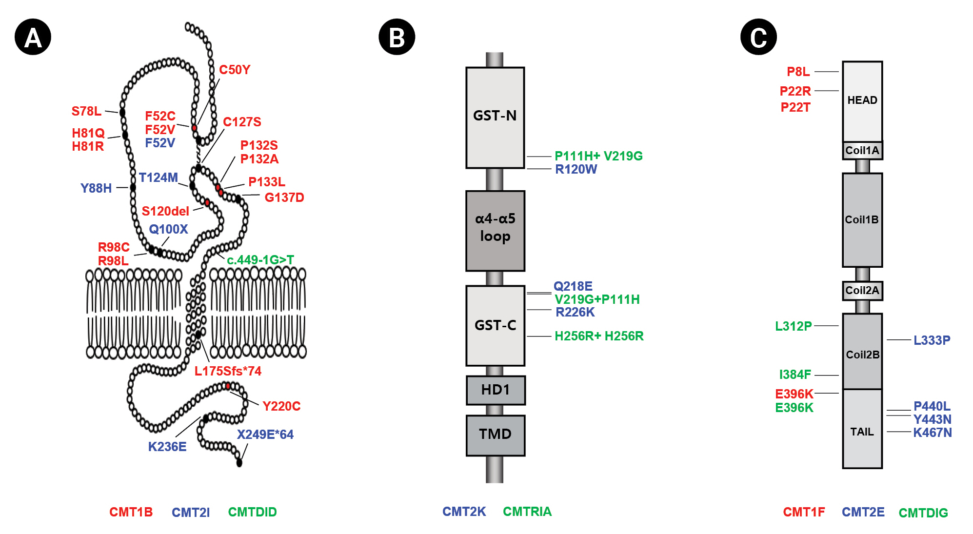
Charcot-Marie-Tooth disease (CMT) is an inherited peripheral neuropathy that is genetically highly heterogeneous, with more than 140 different genes involved [1-3]. Classically, CMT can be divided according to clinical, histopathological, and electrophysiological findings into three types: the demyelinating type (CMT1), with a median motor nerve conduction velocity (MMNCV) below 38 m/s; axonal neuropathy (CMT2), with an MMNCV above 38 m/s; and the intermediate type (CMTDID), with an MMNCV lying between 25 and 45 m/s and nerve pathology showing axonal and/or demyelinating features [4]. Mutations in the myelin protein zero (MPZ), ganglioside-induced differentiation related protein 1 (GDAP1) and neurofilament light-chain polypeptide gene (NEFL) genes have been reported to cause all three CMT types (demyelinating, axonal, and intermediate) (Fig. 1).
Strictly expressed in myelinated Schwann cells, MPZ is a transmembrane protein that is a major component of peripheral myelin [5]. Mutations in MPZ have been reported to cause demyelinating CMT1B (MIM 118200), axonal CMT2I (MIM 607677), and intermediate CMTDID (MIM 607791) (Table 1) [6-11]. CMT1B patients generally exhibit an early onset, while CMT2I patients are characterized by a late onset [12,13]. MPZ-related CMT patients display a spectrum of diverse phenotypes, with phenotypic variations even in the same mutation. [8,14,15].
GDAP1 is mainly expressed in neurons, is located in the outer membrane of the mitochondria, and belongs to the glutathione S-transferase family [16]. Mutations in the GDAP1 gene were reported for the first time in 2002 to cause autosomal recessive (AR) CMT4A (MIM 214400) in Tunisian families [17]. Since then, GDAP1 mutations have been reported to cause axonal forms (CMT2K; MIM 607831) [18,19], axonal forms with vocal cord paresis (MIM 607706) [18] and intermediate forms (CMTRIA; MIM 608340) of disease (Table 1) [20]. GDAP1-related patients harboring AR inheritance exhibit severe clinical features with early onset, but autosomal dominant GDAP1 mutations show mild clinical symptoms with an adult onset [17,18].
NEFL is the most abundant of the three neurofilament proteins, which are major components of the axoskeleton that provide structural support for axons and regulate axon diameter. Patients with NEFL mutations exhibit a diverse phenotypic spectrum [21]. A mutation in NEFL was first reported to cause CMT2E (MIM 607684) in 2000, and several mutations were subsequently revealed to be associated with CMT1F (MIM 607734), CMTDIG (MIM 617882) (Table 1) [21-25].
Most CMT-related genes cause one CMT neuropathy subtype—demyelinating, axonal, or intermediate neuropathy. Thus, it is noteworthy when a single gene causes multiple subtypes of CMT. In this review, we introduce the various types of CMT in a Korean cohort, caused by mutations in MPZ, GDAP1, and NEFL, and describe their clinical, electrophysiological, and genetic characteristics.

Myelin Protein Zero (MPZ)

1) Clinical diversity of MPZ-related patients

The frequency of Korean CMT families with the MPZ mutation was found to be 3.2% in all independent patients and 4.7% in CMT families without PMP22 duplicates (Table 2) [21,23,26-43]. These mutation frequencies were similar to those reported in China (3.3% and 6.4%, respectively) and Britain (3.1% and 5.1%, respectively) but lower than those reported for most other investigated ethnic groups [26,27].
The mean age at onset was 9.3 ± 10.7 years for CMT1B patients, 21.2 ± 13.7 years for CMTDID patients, and 38.7 ± 13.6 years for CMT2I patients (Table 3). The age at onset was significantly different between CMT1B and CMTDID patients (p = 0.025) or CMT2I patients (p < 0.001). However, the age at onset was not significantly different between CMTDID and CMT2I patients. Functional disability was significantly more severe in CMT1B patients than in CMT2I patients (CMT neuropathy score version 2 [CMTNS], p = 0.004, and functional disability scale [FDS], p = 0.022). The CMTNS and FDS values of the CMT1B patients were higher than those of the CMTDID patients, but the difference was not significant. When comparing the degree of disability based on CMTNS, most patients had moderate disease (53%), followed by those with severe disease (31%) and mild disease (17%) in CMT1B families. In CMTDID and CMT2I families, most patients had mild disease (75% and 80%, respectively).

2) Electrophysiological findings in MPZ-related patients

The mean MNCV of CMT1B patients was 12.2 ± 11.0 m/s, which was significantly lower than that of CMT2I patients (46.0 ± 6.6 m/s, p < 0.001) and CMTDID patients (41.3 ± 3.1 m/s, p < 0.001) (Table 3). The mean sensory nerve conduction velocity (SNCV) (7.4 ± 11.7 m/s) of CMT1B patients was significantly lower than that of CMTDID patients (18.0 ± 25.5 m/s, p = 0.021) and CMT2I patients (34.4 ± 4.3 m/s, p < 0.001). In addition, the peroneal MNCV and sural SNCV were significantly reduced in CMT1B patients compared to CMTDID or CMT2I patients. The median motor nerve compound muscle action potential (CMAP) amplitudes (6.0±5.6 mV) in the CMT1B patients were significantly lower in CMTDID (12.8±1.9 mV, p = 0.033) and CMT2I patients (13.0±4.3 mV, p = 0.011). The peroneal nerve CMAP and median and sural sensory nerve action potential (SNAP) amplitudes were also significantly lower in CMT1B patients than in CMTDID and CMT2I patients.

Ganglioside-Induced Differentiation Related Protein 1 (GDAP1)

1) Clinical diversity of GDAP1 mutations

The GDAP1 mutation frequency rate was found to be 0.7% in all patients and 1.0% in patients negative for PMP22 duplication (Table 2). Similar frequencies have been reported in most Asian and Western countries, including Japan, China, Germany, the United States, and the United Kingdom [27-31]. However, higher frequencies of GDAP1 mutations have been reported in certain regions in Italy and Spain [32,33].
The functional disabilities and clinical characteristics were different between CMT2K and CMTRIA patients. CMT2K patients exhibited mild to moderate disabilities, with a late age at onset (19.7 ± 7.7 years), but CMTRIA patients showed severe disabilities with an early age at onset (1.7 ± 0.6 years) (Table 4). Functional disability was significantly more severe in CMTRIA patients than in CMT2K patients. The mean value of the FDS [44] was 2.3 ± 1.4 in CMT2K patients and 5.3 ± 1.2 in CMTRIA patients (p = 0.010). The mean CMTNS score [45] was 11.1±6.6 in CMT2K patients and 24.7 ± 3.2 in CMTRIA patients (p = 0.011). High values of the FDS (scores of 6-7) were observed only in CMTRIA patients. In contrast, low values of the FDS (scores < 5) were observed in CMT2K patients. All three CMTRIA patients were classified in the severe category (CMTNS ≥ 21). Foot deformities were frequent, and four patients had scoliosis. However, no wheelchair dependence, diaphragmatic weakness, vocal cord paresis, or hoarseness was observed.

2) Electrophysiological findings in GDAP1-related patients

Electrophysiological findings verified that CMTRIA patients were more severely affected than CMT2K patients. In CMT2K patients, the conduction velocity mostly did not decrease, excluding nerves with a very low amplitude. In CMTRIA patients, the decreases in CMAP and SNAP amplitudes were even more pronounced, and these parameters were not measured when nerves were explored. These results were worse in the lower extremities than in the upper extremities.

Neurofilament Light-Chain Polypeptide (NEFL)

1) Clinical diversity of NEFL mutations

The frequency of NEFL mutations was reported to range from 0.9% to 2.3% in Japanese and Chinese cohorts and in Korea (Table 2). Data on the frequency of NEFL mutations are extremely limited, though the proportion of the NEFL mutation in CMT has rarely been reported to be below 1%. Therefore, the frequency of NEFL mutations observed in East Asian countries is higher than that reported in other countries.
The prevalence of NEFL mutations was 0.44% in CMT1F patients (5/1,137), 0.35% in CMT2E patients (4/1,137), and 0.70% in CMTDIG patients (8/1,137). The age of onset was thus significantly earlier in CMT1F patients (10.2 ± 7.3 years, p = 0.013) and CMTDIG patients (12.7 ± 7.9, p = 0.007) than in CMT2E patients (24.2 ± 9.4 years) (Table 5). However, the CMTNS and FDS, as measures of disease-related disability, showed no differences among the NEFL-related CMT subtypes. Gait ataxia was identified as the most frequent symptom of NEFL-related CMT patients (78% of CMT1F patients, 50% of CMT2E patients, and 79% of CMTDIG patients). Patients were genetically tested for spinocerebellar ataxia, but no associated mutations were found. In CMT1F and CMTDIG patients, early-onset dementia was observed. Interestingly, ptosis was predominantly observed in CMT2E patients (50%).

2) Electrophysiological findings in NEFL-related patients

In CMT1F patients, the amplitudes of evoked peroneal motor responses were often markedly decreased, and the amplitudes were predominantly unrecordable in 6 of 8 patients (75%) (Table 5). However, peroneal CMAP amplitudes in CMT2E patients could not be recorded in only 1 of 4 patients (25%). Interestingly, peroneal CMAP amplitudes in CMTDIG patients (36%) occupied an intermediate position between the CMT1F and CMT2E patients. The mean MNCV of the median nerve was 16.1 ± 10.5 m/s in CMT1F patients and 47.7 ± 8.1 m/s in CMT2E patients. The mean MNCV of the median nerve was 39.6 ± 4.4 m/s in CMTDIG patients . No SNAP amplitudes of the median nerve were observed in 63% of CMT1F patients, 32% of CMTDIG patients, and 25% of CMT2E patients. Furthermore, sural SNAP amplitudes were not evoked in any of the CMT1F patients, nor were they observed in 36% of CMTDIG patients and 25% of CMT2E patients.

Conclusion

In this review, we described the clinical, electrophysiological, and genetic characteristics of various types of CMT caused by mutations in MPZ, GDAP1, and NEFL. CMT is a peripheral neuropathy with extreme clinical and genetic heterogeneity. Of the 140 causative genes for CMT and other related diseases, MPZ, GDAP1, and NEFL are the only genes that cause all three subtypes of CMT (demyelinating, axonal, and intermediate). CMT is presently incurable; however, the ongoing attempts to treat it with various drugs, dietary supplements, and increase the importance of an exact genetic diagnosis for precision medicine. Therefore, it is important to compare the genetic and clinical features of patients with MPZ, GDAP1, and NEFL mutations. Taken together, a comparison of the causative mutations and clinical features of patients with MPZ-, GDAP1-, and NEFL-related CMT will be the first step in understanding how the different types of CMT are caused, and will enable molecular genetic diagnosis.

Conflict of Interest

No potential conflict of interest relevant to this article was re¬ported.

Fig. 1.
Structure and distribution of mutations in MPZ (A), GDAP1 (B), and NEFL (C) found in Korean CMT families. Vertical arrows indicate the mutation sites. Amino acid changes are indicated in red (demyelinating), blue (axonal), and green (intermediate).
jend-2022-00143f1.jpg
Table 1.
Diseases Caused by Mutations in the MPZ, GDAP1, and NEFL Genes
Gene Locus Phenotype MIM number Heredity
MPZ 1q23.3 CMT1B 118200 AD
CMT2I 607677 AD
CMTDID 607791 AD
GDAP1 8q21.11 CMT4A 214400 AR
CMT2K 607831 AD, AR
CMTRIA 608340 AR
NEFL 8p21.2 CMT1F 607734 AD, AR
CMT2E 607684 AD
CMTDIG 617882 AD

AD, autosomal dominant; AR, autosomal recessive.

Table 2.
MPZ, GDAP1, and NEFL Mutation Detection Rates in Various Populations
Gene Population Frequency
Reference
Total CMT patients (%) CMT patients excluding CMT1A (%)
MPZ Korean 3.2 4.7 [34]
Chinese 3.3 6.4 [26]
Japanese 5.1 NA [29]
German 4.2 6.4 [31]
British 3.1 5.1 [27]
American 4.1 6.5 [35]
Spanish 4.3 7.5 [32]
Italian 4.3 12.3 [33]
Hungarian 4.5 7.5 [36]
Norwegian 6.0 NA [37]
Russian 3.5 5.2 [21]
Finnish 5.2 NA [38]
Austrian 4.0 NA [39]
GDAP1 Korean 0.7 1.0 [40]
Chinese NA 1.6 [30]
Japanese NA 0.9 [29]
British 0.5 0.8 [27]
American 0.7 1.6 [28]
Spanish 11.1 20.7 [32]
Italian 5.4 11.0 [33]
NEFL Korean 1.5 2.1 [41]
Chinese 1.9 3.7 [26]
Japanese 0.9 NA [29]
2.3 2.7 [23]
German 0.0 0.0 [31]
0.1 0.1 [42]
British 0.2 0.2 [27]
American 0.7 1.6 [28]
0.7 1.1 [35]
0.5 0.8 [43]
Spanish 0.9 1.6 [32]
Italian 0.6 1.8 [33]
Norwegian 0.7 NA [37]

CMT, Charcot-Marie-Tooth disease; NA, not available.

Table 3.
Clinical and Electrophysiological Features of Korean CMT Patients with MPZ Mutations
Item CMT1B CMT2I CMTDID p-value
1B vs. 2I 1B vs. DID 2I vs. DID ANOVA
Patient number 48 7 5
Female ratio (%) 54 14 20
Examined age (y) 26.4 ± 19.6 51.1 ± 11.7 40.8 ± 23.3 0.002 0.129 0.332 0.005
Onset age (y) 9.3 ± 10.7 38.7 ± 13.6 21.2 ± 13.7 < 0.001 0.025 0.053 < 0.001
Disability score
 CMTNS 16.0 ± 7.4 8.0 ± 3.2 9.8 ± 1.0 0.004 0.102 0.326 0.023
 FDS 2.8 ± 1.3 1.3 ± 0.5 1.8 ± 0.4 0.022 0.092 0.092 0.004
Nerve conduction studies
Patient number 38 5 4
 Median motor nerve
  CMAP (mV) 6.0 ± 5.6 13.0 ± 4.3 12.8 ± 1.9 0.011 0.033 0.915 0.006
  MNCV (m/s) 12.2 ± 11.0 46.0 ± 6.6 41.3 ± 3.1 < 0.001 < 0.001 0.431 < 0.001
 Peroneal nerve
  CMAP (mV) 0.9 ± 2.0 2.1 ± 2.8 4.0 ± 3.1 0.231 0.001 0.132 0.002
  MNCV (m/s) 5.1 ± 9.0 20.4 ± 19.4 25.7 ± 17.2 0.004 < 0.001 0.278 < 0.001
 Median sensory nerve
  SNAP (μV) 2.3 ± 4.7 15.6 ± 12.5 10.8 ± 15.3 < 0.001 < 0.001 0.718 < 0.001
  SNCV (m/s) 7.4 ± 11.7 34.4 ± 4.3 18.0 ± 25.5 < 0.001 0.021 0.78 < 0.001
 Sural nerve
  SNAP (μV) 1.1 ± 3.5 5.4 ± 5.9 6.9 ± 9.7 0.059 0.001 0.346 0.002
  SNCV (m/s) 3.4 ± 9.0 17.2 ± 14.9 13.3 ± 18.7 0.02 0.016 0.643 0.007

All data are expressed as the mean ± standard deviation. Normal nerve conduction velocity values: motor median nerve ≥ 50.5 m/s; sensory median nerve ≥ 39.3 m/s; sural nerve ≥ 32.1 m/s. Normal amplitude values: motor median nerve ≥ 6 mV; sensory median nerve ≥ 8.8 μV; sural nerve ≥ 6.0 μV.

CMT, Charcot-Marie-Tooth disease; ANOVA, analysis of variance; CMTNS, CMT neuropathy score; FDS, functional disability scale; CMAP, compound muscle action potential; MNCV, motor nerve conduction velocity; SNAP, sensory nerve action potential; SNCV, sensory nerve conduction velocity.

Table 4.
Clinical and Electrophysiological Features of Korean CMT Patients with GDAP1 Mutations
Item CMT2K CMTRIA p-value
Patient number 7 3
Female ratio (%) 29 67
Examined age (y) 42.6 ± 15.7 9.7 ± 4.2 0.009
Onset age (y) 19.7 ± 7.7 1.7 ± 0.6 0.004
Disability score
 CMTNS 11.1 ± 6.6 24.7 ± 3.2 0.011
 FDS 2.3 ± 1.4 5.3 ± 1.2 0.010
Nerve conduction studies
 Patient number 7 3
  Median motor nerve
   CMAP (mV) 11.2 ± 5.8 1.4 ± 1.2 0.024
   MNCV (m/s) 52.0 ± 5.8 32.3 ± 28.0 0.092
  Peroneal nerve
   CMAP (mV) 1.8 ± 1.8 0.0 ± 0.1 0.127
   MNCV (m/s) 21.7 ± 20.4 0.0 ± 0.0 0.113
  Median sensory nerve
   SNAP (μV) 6.3 ± 3.0 0.9 ± 1.3 0.051
   SNCV (m/s) 38.2 ± 1.2 12.5 ± 17.7 0.005
  Sural nerve
   SNAP (μV) 0.2 ± 0.6 0.0 ± 0.0 0.604
   SNCV (m/s) 4.8 ± 11.8 0.0 ± 0.0 0.604

All data are expressed as the mean ± standard deviation. Normal nerve conduction velocity values: motor median nerve ≥ 50.5 m/s; sensory median nerve ≥ 39.3 m/s; sural nerve ≥ 32.1 m/s. Normal amplitude values: motor median nerve ≥ 6 mV; sensory median nerve ≥ 8.8 μV; sural nerve ≥ 6.0 μV.

CMT, Charcot-Marie-Tooth disease; ANOVA, analysis of variance; CMTNS, CMT neuropathy score; FDS, functional disability scale; CMAP, compound muscle action potential; MNCV, motor nerve conduction velocity; SNAP, sensory nerve action potential; SNCV, sensory nerve conduction velocity.

Table 5.
Clinical and Electrophysiological Features of Korean CMT Patients with NEFL Mutations
Item CMT1F CMT2E CMTDIG p-value
1B vs. 2I 1B vs. DIG 2I vs. DIG ANOVA
Patient number 9 4 24
Female ratio (%) 44 25 50
Examined age (y) 32.4 ± 16.8 42.7 ± 6.9 37.3 ± 16.3 0.271 0.453 0.524 0.536
Onset age (y) 10.2 ± 7.3 24.2 ± 9.4 12.1 ± 7.4 0.013 0.523 0.007 0.011
Disability score
 CMTNS 24.1 ± 5.1 16.2 ± 12.3 18.4 ± 7.9 0.136 0.071 0.641 0.169
 FDS 4.9 ± 1.9 3.2 ± 3.0 3.4 ± 2.0 0.243 0.055 0.914 0.165
Nerve conduction studies
 Patient number 8 4 22
  Median motor nerve
   CMAP (mV) 3.1 ± 4.3 8.7 ± 5.2 9.6 ± 3.5 0.075 < 0.001 0.666 0.001
   MNCV (m/s) 16.1 ± 10.5 47.7 ± 8.1 39.6 ± 4.4 < 0.001 < 0.001 0.007 < 0.001
  Peroneal nerve
   CMAP (mV) 0.5 ± 1.0 3.1 ± 4.0 1.7 ± 2.0 0.104 0.122 0.298 0.148
   MNCV (m/s) 6.4 ± 12.2 26.3 ± 18.9 21.2 ± 17.1 0.049 0.033 0.591 0.066
  Median sensory nerve
   SNAP (μV) 4.6 ± 7.8 9.7 ± 15.4 5.0 ± 6.0 0.449 0.877 0.275 0.509
   SNCV (m/s) 13.0 ± 18.5 29.0 ± 19.4 23.7 ± 17 0.194 0.146 0.581 0.246
  Sural nerve
   SNAP (μV) 0.0 ± 0.0 6.0 ± 9.0 2.5 ± 2.6 0.073 0.011 0.119 0.028
   SNCV (m/s) 0.0 ± 0.0 23.4 ± 16.4 18.2 ± 14.6 0.002 0.002 0.522 0.004

All data are expressed as the mean ± standard deviation. Normal nerve conduction velocity values: motor median nerve ≥ 50.5 m/s; sensory median nerve ≥ 39.3 m/s; sural nerve ≥ 32.1 m/s. Normal amplitude values: motor median nerve ≥ 6 mV; sensory median nerve ≥ 8.8 μV; sural nerve ≥ 6.0 μV.

CMT, Charcot-Marie-Tooth disease; ANOVA, analysis of variance; CMTNS, CMT neuropathy score; FDS, functional disability scale; CMAP, compound muscle action potential; MNCV, motor nerve conduction velocity; SNAP, sensory nerve action potential; SNCV, sensory nerve conduction velocity.

References

1. Madrid R, Bradley WG, Davis CJ: The peroneal muscular atrophy syndrome: clinical, genetic, electrophysiological and nerve biopsy studies. Part 2. Observations on pathological changes in sural nerve biopsies. J Neurol Sci 1977;32:91-122.
crossref pmid pmc
2. Cortese A, Wilcox JE, Polke JM, Poh R, Skorupinska M, Rossor AM, et al: Targeted next-generation sequencing panels in the diagnosis of Charcot-Marie-Tooth disease. Neurology 2020;94:e51-e61.
crossref pmid pdf
3. Gonzaga-Jauregui C, Harel T, Gambin T, Kousi M, Griffin LB, Francescatto L, et al: Exome sequence analysis suggests that genetic burden contributes to phenotypic variability and complex neuropathy. Cell Rep 2015;12:1169-1183.
crossref pmid
4. Rossor AM, Polke JM, Houlden H, Reilly MM: Clinical implications of genetic advances in Charcot-Marie-Tooth disease. Nat Rev Neurol 2013;9:562-571.
crossref pmid
5. Lemke G, Axel R: Isolation and sequence of a cDNA encoding the major structural protein of peripheral myelin. Cell 1985;40:501-508.
crossref pmid pdf
6. De Jonghe P, Timmerman V, Ceuterick C, Nelis E, De Vriendt E, Löfgren A, et al: The Thr124Met mutation in the peripheral myelin protein zero (MPZ) gene is associated with a clinically distinct Charcot-Marie-Tooth phenotype. Brain 1999;122(Pt 2):281-290.
pmid
7. Hayasaka K, Himoro M, Sato W, Takada G, Uyemura K, Shimizu N, et al: Charcot-Marie-Tooth neuropathy type 1B is associated with mutations of the myelin P0 gene. Nat Genet 1993;5:31-34.
crossref pmid pmc
8. Kochański A: Mutations in the Myelin Protein Zero result in a spectrum of Charcot-Marie-Tooth phenotypes. Acta Myol 2004;23:6-9.
crossref pmid pmc
9. Mastaglia FL, Nowak KJ, Stell R, Phillips BA, Edmondston JE, Dorosz SM, et al: Novel mutation in the myelin protein zero gene in a family with intermediate hereditary motor and sensory neuropathy. J Neurol Neurosurg Psychiatry 1999;67:174-179.
crossref pmid
10. Sanmaneechai O, Feely S, Scherer SS, Herrmann DN, Burns J, Muntoni F, et al: Genotype-phenotype characteristics and baseline natural history of heritable neuropathies caused by mutations in the MPZ gene. Brain 2015;138(Pt 11):3180-3192.
crossref pmid
11. Senderek J, Hermanns B, Lehmann U, Bergmann C, Marx G, Kabus C, et al: Charcot-Marie-Tooth neuropathy type 2 and P0 point mutations: two novel amino acid substitutions (Asp61Gly; Tyr119Cys) and a possible “hotspot” on Thr124Met. Brain Pathol 2000;10:235-248.
crossref pmid
12. Hattori N, Yamamoto M, Yoshihara T, Koike H, Nakagawa M, Yoshikawa H, et al: Demyelinating and axonal features of Charcot-Marie-Tooth disease with mutations of myelin-related proteins (PMP22, MPZ and Cx32): a clinicopathological study of 205 Japanese patients. Brain 2003;126(Pt 1):134-151.
crossref pmid
13. Shy ME, Jáni A, Krajewski K, Grandis M, Lewis RA, Li J, et al: Phenotypic clustering in MPZ mutations. Brain 2004;127(Pt 2):371-384.
crossref pmid
14. Mazzeo A, Muglia M, Rodolico C, Toscano A, Patitucci A, Quattrone A, et al: Charcot-Marie-Tooth disease type 1B: marked phenotypic variation of the Ser78Leu mutation in five Italian families. Acta Neurol Scand 2008;118:328-332.
crossref pmid pmc
15. Senderek J, Ramaekers VT, Zerres K, Rudnik-Schöneborn S, Schröder JM, Bergmann C: Phenotypic variation of a novel nonsense mutation in the P0 intracellular domain. J Neurol Sci 2001;192:49-51.
crossref pmid pdf
16. Estela A, Pla-Martín D, Sánchez-Piris M, Sesaki H, Palau F: Charcot-Marie-Tooth-related gene GDAP1 complements cell cycle delay at G2/M phase in Saccharomyces cerevisiae fis1 gene-defective cells. J Biol Chem 2011;286:36777-36786.
crossref pmid pdf
17. Baxter RV, Ben Othmane K, Rochelle JM, Stajich JE, Hulette C, Dew-Knight S, et al: Ganglioside-induced differentiation-associated protein-1 is mutant in Charcot-Marie-Tooth disease type 4A/8q21. Nat Genet 2002;30:21-22.
crossref pmid pmc
18. Cuesta A, Pedrola L, Sevilla T, García-Planells J, Chumillas MJ, Mayordomo F, et al: The gene encoding ganglioside-induced differentiation-associated protein 1 is mutated in axonal Charcot-Marie-Tooth type 4A disease. Nat Genet 2002;30:22-25.
crossref pmid
19. Claramunt R, Pedrola L, Sevilla T, López de Munain A, Berciano J, Cuesta A, et al: Genetics of Charcot-Marie-Tooth disease type 4A: mutations, inheritance, phenotypic variability, and founder effect. J Med Genet 2005;42:358-365.
crossref pmid pmc
20. Senderek J, Bergmann C, Ramaekers VT, Nelis E, Bernert G, Makowski A, et al: Mutations in the ganglioside-induced differentiation-associated protein-1 (GDAP1) gene in intermediate type autosomal recessive Charcot-Marie-Tooth neuropathy. Brain 2003;126(Pt 3):642-649.
crossref pmid
21. Mersiyanova IV, Perepelov AV, Polyakov AV, Sitnikov VF, Dadali EL, Oparin RB, et al: A new variant of Charcot-Marie-Tooth disease type 2 is probably the result of a mutation in the neurofilament-light gene. Am J Hum Genet 2000;67:37-46.
crossref pmid pdf
22. Jordanova A, De Jonghe P, Boerkoel CF, Takashima H, De Vriendt E, Ceuterick C, et al: Mutations in the neurofilament light chain gene (NEFL) cause early onset severe Charcot-Marie-Tooth disease. Brain 2003;126(Pt 3):590-597.
crossref pmid pmc
23. Abe A, Numakura C, Saito K, Koide H, Oka N, Honma A, et al: Neurofilament light chain polypeptide gene mutations in Charcot-Marie-Tooth disease: nonsense mutation probably causes a recessive phenotype. J Hum Genet 2009;54:94-97.
crossref pmid pdf
24. Yum SW, Zhang J, Mo K, Li J, Scherer SS: A novel recessive Nefl mutation causes a severe, early-onset axonal neuropathy. Ann Neurol 2009;66:759-770.
crossref pmid pmc pdf
25. Berciano J, García A, Peeters K, Gallardo E, De Vriendt E, Pelayo-Negro AL, et al: NEFL E396K mutation is associated with a novel dominant intermediate Charcot-Marie-Tooth disease phenotype. J Neurol 2015;262:1289-1300.
crossref pmid
26. Hsu YH, Lin KP, Guo YC, Tsai YS, Liao YC, Lee YC: Mutation spectrum of Charcot-Marie-Tooth disease among the Han Chinese in Taiwan. Ann Clin Transl Neurol 2019;6:1090-1101.
crossref pmid pmc pdf
27. Murphy SM, Laura M, Fawcett K, Pandraud A, Liu YT, Davidson GL, et al: Charcot-Marie-Tooth disease: frequency of genetic subtypes and guidelines for genetic testing. J Neurol Neurosurg Psychiatry 2012;83:706-710.
crossref pmid
28. DiVincenzo C, Elzinga CD, Medeiros AC, Karbassi I, Jones JR, Evans MC, et al: The allelic spectrum of Charcot-Marie-Tooth disease in over 17,000 individuals with neuropathy. Mol Genet Genomic Med 2014;2:522-529.
crossref pmid pdf
29. Yoshimura A, Yuan JH, Hashiguchi A, Ando M, Higuchi Y, Nakamura T, et al: Genetic profile and onset features of 1005 patients with Charcot-Marie-Tooth disease in Japan. J Neurol Neurosurg Psychiatry 2019;90:195-202.
crossref pmid
30. Pakhrin PS, Xie Y, Hu Z, Li X, Liu L, Huang S, et al: Genotype-phenotype correlation and frequency of distribution in a cohort of Chinese Charcot-Marie-Tooth patients associated with GDAP1 mutations. J Neurol 2018;265:637-646.
crossref pmid pmc
31. Gess B, Schirmacher A, Boentert M, Young P: Charcot-Marie-Tooth disease: frequency of genetic subtypes in a German neuromuscular center population. Neuromuscul Disord 2013;23:647-651.
crossref pmid pdf
32. Sivera R, Sevilla T, Vílchez JJ, Martínez-Rubio D, Chumillas MJ, Vázquez JF, et al: Charcot-Marie-Tooth disease: genetic and clinical spectrum in a Spanish clinical series. Neurology 2013;81:1617-1625.
crossref pmid
33. Sadjadi R, Reilly MM, Shy ME, Pareyson D, Laura M, Murphy S, et al: Psychometrics evaluation of Charcot-Marie-Tooth Neuropathy Score (CMTNSv2) second version, using Rasch analysis. J Peripher Nerv Syst 2014;19:192-196.
crossref pmid pmc pdf
34. Kim HJ, Nam SH, Kwon HM, Lim SO, Park JH, Kim HS, et al: Genetic and clinical spectrums in Korean Charcot-Marie-Tooth disease patients with myelin protein zero mutations. Mol Genet Genomic Med 2021;9:e1678.
crossref pmid pmc pdf
35. Fridman V, Bundy B, Reilly MM, Pareyson D, Bacon C, Burns J, et al: CMT subtypes and disease burden in patients enrolled in the Inherited Neuropathies Consortium natural history study: a cross-sectional analysis. J Neurol Neurosurg Psychiatry 2015;86:873-878.
crossref pmid
36. Milley GM, Varga ET, Grosz Z, Nemes C, Arányi Z, Boczán J, et al: Genotypic and phenotypic spectrum of the most common causative genes of Charcot-Marie-Tooth disease in Hungarian patients. Neuromuscul Disord 2018;28:38-43.
crossref pmid
37. Østern R, Fagerheim T, Hjellnes H, Nygård B, Mellgren SI, Nilssen Ø: Diagnostic laboratory testing for Charcot Marie Tooth disease (CMT): the spectrum of gene defects in Norwegian patients with CMT and its implications for future genetic test strategies. BMC Med Genet 2013;14:94.
crossref pmid pmc pdf
38. Silander K, Meretoja P, Juvonen V, Ignatius J, Pihko H, Saarinen A, et al: Spectrum of mutations in Finnish patients with Charcot-Marie-Tooth disease and related neuropathies. Hum Mutat 1998;12:59-68.
crossref pmid
39. Miltenberger-Miltenyi G, Schwarzbraun T, Löscher WN, Wanschitz J, Windpassinger C, Duba HC, et al: Identification and in silico analysis of 14 novel GJB1, MPZ and PMP22 gene mutations. Eur J Hum Genet 2009;17:1154-1159.
crossref pmid pmc pdf
40. Kim HS, Kim HJ, Nam SH, Kim SB, Choi YJ, Lee KS, et al: Clinical and neuroimaging features in Charcot-Marie-Tooth patients with GDAP1 mutations. J Clin Neurol 2021;17:52-62.
crossref pmid pdf
41. Kim HJ, Kim SB, Kim HS, Kwon HM, Park JH, Lee AJ, et al: Phenotypic heterogeneity in patients with NEFL-related Charcot-Marie-Tooth disease. Mol Genet Genomic Med 2022;10:e1870.
crossref pmid pmc pdf
42. Rudnik-Schöneborn S, Tölle D, Senderek J, Eggermann K, Elbracht M, Kornak U, et al: Diagnostic algorithms in Charcot-Marie-Tooth neuropathies: experiences from a German genetic laboratory on the basis of 1206 index patients. Clin Genet 2016;89:34-43.
crossref pmid pdf
43. Saporta AS, Sottile SL, Miller LJ, Feely SM, Siskind CE, Shy ME: Charcot-Marie-Tooth disease subtypes and genetic testing strategies. Ann Neurol 2011;69:22-33.
crossref pmid pmc
TOOLS
Share :
Facebook Twitter Linked In Google+ Line it
METRICS Graph View
  • 0 Crossref
  •     Scopus
  • 1,650 View
  • 12 Download
Related articles in J Korean Assoc EMG Electrodiagn Med


ABOUT
ARTICLE CATEGORY

Browse all articles >

BROWSE ARTICLES
EDITORIAL POLICY
FOR CONTRIBUTORS
Editorial Office
Department of Physical Medicine and Rehabilitation, Korea University Ansan Hospital
123 Jeokgeum-ro, Danwon-gu, Ansan 15355, Korea
Tel: +82-31-412-5330    Fax: +82-31-412-4215    E-mail: editjend@gmail.com                

Copyright © 2024 by Korean Association of EMG Electrodiagnostic Medicine.

Developed in M2PI

Close layer
prev next