Introduction
Vertebral osteomyelitis (VO) is a rare disease that is characterized by infection of the vertebral body and adjacent intervertebral discs [1-3]. VO may cause neurological complications, such as motor weakness or sensory abnormalities [1,3,4].
Epidural block, which is commonly used in patients with radicular pain, allows the effective delivery of drugs to the epiradicular space. Although epidural block is generally known to be a safe method, complications including infection occasionally occur [5-7].
Herein, we present a rare case of VO with serious neuromuscular complications, including cervical and lumbar radiculopathy, myelopathy, and multiple pyogenic abscesses, which developed in a patient who underwent lumbar transforaminal epidural block for back pain.
It is difficult for a physician to determine the exact cause of weakness when multiple etiologies are suspected. Electrodiagnostic studies such as nerve conduction studies (NCS) and needle electromyography (EMG) could enhance the differential diagnosis of the etiology. The use of electrodiagnostic studies in addition to imaging studies, such as computed tomography (CT) and magnetic resonance imaging (MRI), helped to determine the degree and locations of nerve and muscle damage and decide on a treatment strategy.
Furthermore, the early application of a multidisciplinary approach, including medical treatment, surgery, and comprehensive rehabilitation enhanced the patient's prognosis and functional recovery.
Case Report
A 64-year-old man visited an outpatient clinic due to low back pain radiating to the left lower extremity that started 1 month ago. A neurological examination revealed no sensory deficit or motor weakness. MRI revealed herniation of the left L4-5 intervertebral disc, and the patient underwent transforaminal epidural block on the left side at the L5 level. However, 2 days after the procedure, he developed general weakness. Since the symptoms worsened, he was admitted to the emergency department 5 days after the procedure.
The patient had been diagnosed with diabetes mellitus (DM) 10 years ago. He had been taking medication intermittently in the last year and stopped taking it entirely in the last 2 weeks.
He complained of generalized weakness in the bilateral upper and lower extremities. In a manual muscle strength evaluation, the bilateral upper limb strength was measured as Medical Research Council (MRC) grade 4-5/5 and bilateral lower limb strength as MRC grade 2-3/5.
In a sensory evaluation, both light touch and pinprick tests showed no abnormalities. The deep tendon reflex was normal, and there were no pathological reflexes. The sensation of deep anal pressure was decreased and voluntary anal contraction was slightly weak, but the bulbocavernous reflex was preserved.
His white blood cell count was 8,100 cells/┬ĄL with a neutrophil predominance, the serum C- reactive protein level was 43.05 mg/L (normal range, < 0.5 mg/L), and the erythrocyte sedimentation rate was 120 mm/h (normal range, 0-20 mm/h). The procalcitonin level was 5.71 ng/mL (normal range, 0-0.5 ng/mL). The hemoglobin A1C level was 8.4% (normal range, < 6.1%), which was consistent with an elevated estimated average glucose level of 391 mg/dL.
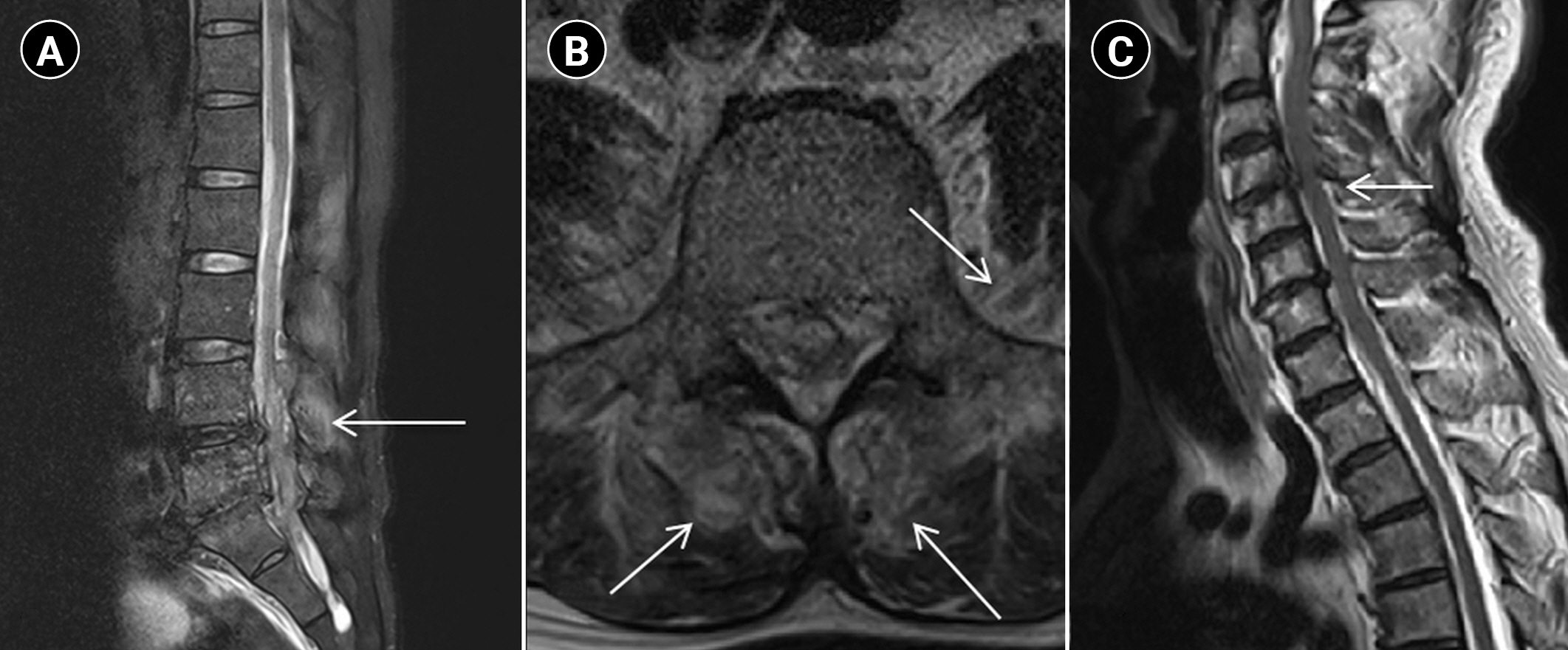
Lumbar spine MRI showed degenerative changes and VO with fluid collection at the L4-5 and L5-S1 levels, as well as abscesses in the bilateral paraspinalis and left psoas muscles. Cervical spine MRI showed degenerative changes at the C4-5 level without any evidence of infection (Fig. 1). Decompressive laminectomy and spinal abscess removal were performed the next day, and methicillin-susceptible Staphylococcus aureus was simultaneously identified in the tissue culture and blood culture.
Intensive antibiotic therapy was started for 2 weeks, but the patientŌĆÖs weakness progressed. A repeat neurological examination was performed. In the sensory evaluation, both light touch and pinprick tests showed sensory deficits below the C3 level. The upper limb weakness worsened to MRC grade 3/5 on the right side and MRC grade 2-3/5 on the left side; therefore, we performed cervical spine MRI and abdominopelvic CT. Cervical spine MRI showed discitis at C4/5 and epidural abscesses at C1-C4 with spinal cord compression (Fig. 2). Abdominopelvic CT showed bilateral abscesses in the iliopsoas, paraspinalis at L4-5, and the right gluteus maximus muscle (Fig. 3). Surgical decompression of the cervical spine and CT-guided aspiration of the abscess in the right gluteus maximus muscle were performed.
Electrodiagnostic studies were conducted to determine the cause of weakness.
A sensory NCS showed delayed-onset latency of sensory nerve action potential in the bilateral median and sural nerves, and a motor NCS showed delayed distal latency of compound muscle action potentials in the bilateral median and tibial nerves, as well as the right ulnar and peroneal nerves.
Somatosensory evoked potentials in both the median and tibial nerves showed delayed response in all extremities. Motor evoked potentials showed more delayed responses in the left upper extremity compared to the right side.
Needle EMG showed increased insertional activity and abnormal spontaneous activity at rest (grades 2 to 3+) in the bilateral biceps, pronator teres, flexor carpi ulnaris, extensor digitorum communis, abductor digiti minimi, abductor pollicis brevis, triceps, deltoid, tibialis anterior, extensor hallucis longus, gastrocnemius, peroneus longus, iliacus, iliopsoas, and paraspinalis (C5-T1, L2-S1) muscles. During minimal muscle contraction, polyphasic motor unit action potentials of large amplitude and long duration were observed, and the recruitment pattern generally decreased during maximum muscle contraction in the above-mentioned muscles (Tables 1, 2). It revealed multiple etiologies of weakness, including cervical and lumbar polyradiculopathy, polyneuropathy, and cervical myelopathy.
Treatment was started with an early multidisciplinary approach. Intravenous antibiotic therapy was applied for 6 additional weeks. Strengthening exercises for both upper and lower extremities with progressive gait training were performed in sessions.
After 2 months of hospitalization, the patientŌĆÖs functional state improved to achieve independent walker-assisted gait.
Discussion
VO is a rare disease that accounts for approximately 1% of all skeletal infections [2,8]. It mainly involves 2 vertebral bodies and the adjacent intervertebral discs [4]. Most cases involve a single region (94%), and the most common location was reported to be the lumbar spine (58%) [3,8]. However, the involvement of multiple levels has rarely been reported in previous studies, accounting for 4% of all VO cases, and only 3% of cases were skip lesions [2].
VO has an insidious onset, and its progression is slow [4]. Neurological deficits, including motor or sensory deficits and bladder or bowel dysfunction, were reported in one-third of cases [3]. S. aureus is the most commonly isolated causative agent (32%-67%) [2]. VO is mostly caused by hematogenous seeding (50%), Direct inoculation after iatrogenic procedures (15%-40%), or contiguous spread from an adjacent area (3%) [1,4]. VO often causes myositis or abscess in surrounding structures, such as the paravertebral, epidural, or psoas abscesses [3,8]. In addition, cervical involvement significantly increases the risk of spinal cord compression, according to Doutchi et al. [8]. The evaluation of VO should include a history, physical examination, laboratory results, and MRI [4]. MRI can diagnose VO with high accuracy (90%).
Epidural injections are a widely used non-surgical treatment for patients with radicular pain by disc herniation. Transforaminal injection is a method that effectively delivers corticosteroids to the epiradicular space [6]. It is generally known to be a safe method, but epidural space infections, discitis, and osteomyelitis can occasionally occur [5-7]. Absolute contraindications include local infection around the injection site, anticoagulant use, uncontrolled DM, and congestive heart failure [6]. In this case, based on the patient's medical history, his DM was uncontrolled for the last 2-3 months. The patient was in an immunosuppressed state, which caused the disseminated infection.
It is important to implement an intensive and early multidisciplinary approach, including medical treatment, surgery, and rehabilitative treatment, to achieve favorable outcomes in VO. In the case presented herein, a rapid diagnosis and a multidisciplinary approach were applied to determine the focus of infection and causative organism, and early surgical and rehabilitative treatment was performed in parallel.
Of particular note, a detailed rehabilitative approach and the identification of multiple causes of paralysis are essential for patients with VO. Since neurologic complications can cause functional loss and low quality of life, the early application of comprehensive rehabilitation could be the key to a good prognosis, the recovery of functional ability, and improvement of the quality of life for patients with VO
Physicians often experience difficulty in the field when a precise differential diagnosis of weakness is necessary. Electrodiagnostic studies are advantageous for determining the degree of damage and for follow-up of improvement in patients with VO. Electrodiagnostic studies could be a good diagnostic tool for physicians in determining the etiology of weakness in patients with VO.
There is a potential risk of iatrogenic complications when performing needle EMG, including bleeding, infection, nerve injury, pneumothorax, and other local trauma [9]. In particular, the risk of seeding infection should be considered in patients with a suspected infection [10]. There is no quantitative evidence on the theoretical risk of seeding infection according to previous studies [10]. To reduce this risk, sterilized, disposable concentric needles were used. The examiner washed hands before and after the procedure, ad patient's skin was cleaned with alcohol. It was clear in our case that a precise diagnosis was more important than the theoretical risk [9,10].